COPD exacerbation management X21 Confirm exacerbation and categorise severity Assessment of severity of the exacerbation includes a medical history examination spirometry and in severe cases FEV1 40 predicted blood gas measurements chest x- rays and electrocardiography. A chest x-ray would be a reasonable study to look for masses infiltrates edema or signs of obstructive airflow suggestive of COPD.
Chronic Obstructive Pulmonary Disease Copd Radiology Key

Acute Exacerbation Of Chronic Obstructive Pulmonary Disease Wikipedia

Ditch X Ray To Diagnose Copd Study Finds
A chest X-ray is not useful to establish a diagnosis of COPD but it is of use in either excluding other conditions or including comorbidities such as pulmonary fibrosis and bronchiectasis.

Copd x ray. An X-ray in COPD may not reveal as much if the condition is primarily chronic bronchitis. A chest X-ray isnt used to diagnose COPD but it may help rule out conditions that cause similar symptoms such as pneumonia. Exercise testing to determine if the oxygen level in your blood drops when you exercise.
Principles of Reading 1. But with emphysema more structural problems of the lungs can be seen on an X-ray. Dark-field X-rays visualize early changes in the alveolar structure caused by the lung disease COPD and require only one 50th of the radiation dose typically applied in X.
Note the larger appearing heart on the AP view. CT scans can also be used to screen for lung cancer. Often a lateral view usually accompanies a PAAP chest X-rayThis can be helpful in settings where the single.
Spirometry is performed before and after administering an. COPD chronic obstructive pulmonary disease. What are the stages of COPD.
The study X-ray dark-field chest imaging for detection and quantification of emphysema in patients with chronic obstructive pulmonary disease. A CT scan of your lungs can help detect emphysema and help determine if you might benefit from surgery for COPD. Chronic Obstructive Pulmonary Disease COPD is a term used to describe chronic lung diseases including emphysema and chronic bronchitis.
COPD can often be prevented. COPD poses a major health and. A chest X-ray may be able to show enlarged lungs that can occur in some patients with COPD due to hyperinflation.
The literature of acute exacerbation of chronic obstructive pulmonary disease COPD is fast expanding. A chest X-ray can show emphysema one of the main causes of COPD. This review focuses on several aspects of acute exacerbation of COPD AECOPD including epidemiology diagnosis and management.
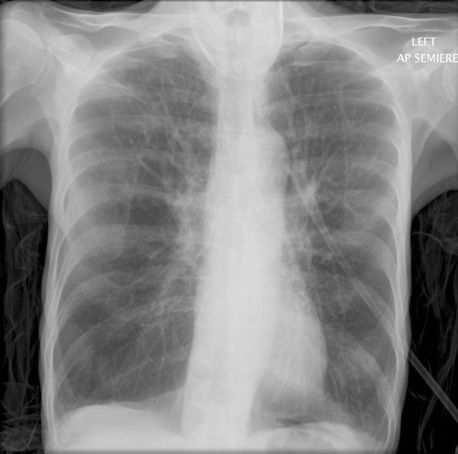
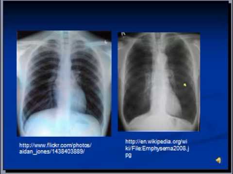
Onset after age 40 Persistence of symptoms despite treatment Abnormal lung function between symptoms. Various tests such as lung function tests a chest x-ray or CT scan and blood tests. COPD bullous emphysema Bullous emphysema manifests on a chest X-ray with areas of low density black with thinning of the pulmonary vessels predominantly affecting the upper zones The lower part of the lungs may appear denser whiter in normal subjects because of overlying breast tissue but in this individual the pulmonary vessels appear normal in this area.
In advanced COPD a chest X. Pulmonary function tests - Essential in the diagnosis staging and monitoring of COPD. Comparison of PA vs.
This disease is characterized by breathlessness. A diagnosis of chronic obstructive pulmonary disease COPD is based on a variety of things. This exam can help support the diagnosis of COPD by producing images of the lungs to evaluate symptoms of shortness of breath or chronic cough.
The resulting image may reveal enlarged lungs a. Chest X-ray or chest CT scan to look for lung changes that are caused by COPD. Your doctor will diagnose COPD based on your signs and symptoms your medical and family histories and test results.
However X-ray is more useful to help rule out or rule in other problems that may cause symptoms similar to COPD such as pneumonia. This is because the distance is increased between the film and the heartallowing for the X-rays to spread for a greater distance before developing the film Lateral views rightleft. The shadows on a chest X-ray test depend on the degree of absorbed radiation by the particular organ based on its composition.
A chest X-ray and other tests may also help zero in on a diagnosis see below. Definition clinical manifestations. Progressive means the disease gets worse over time.
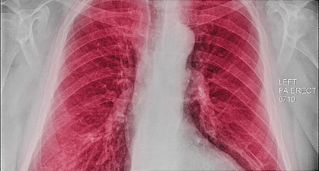
A chest X-ray of someone with suspected chronic obstructive pulmonary disease or COPD is a standard part of a diagnosis. As mentioned earlier the image on the chest X-ray film is in shades of black and white similar to a negative of a regular photograph. A diagnostic accuracy study was published in The Lancet Digital Health.
Characteristic signs of COPD on X-ray include hyperinflation shown by a flattened diaphragm and an increased retrosternal air space and lung hyperlucency. Patients should be provided with and bring a summary of their medical problems and treatment eg a. A CT scan is a type of X-ray that creates a more detailed image than a standard X-ray.
An X-ray can also rule out other lung problems or heart failure. While a chest x-ray may not show COPD until it is severe the images may show enlarged lungs air pockets or a flattened diaphragm. A normal chest x-ray does not rule out COPD.
AP views of chest X-rays. Miscellaneous such as pacemakers catheters etc. A chest X-ray shows COPD signs.
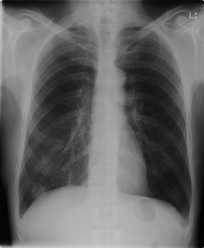
Chronic obstructive pulmonary disease. Normal chest X-ray The primary features of COPD include the following. COPD can cause coughing that produces large amounts of a slimy substance called mucus wheezing shortness of breath chest tightness and other symptoms.
COPD is currently an incurable disease but with the right diagnosis and treatment there are many things you can do to breathe better and enjoy life and live for many years. COPD or chronic obstructive pulmonary disease is a progressive disease that makes it hard to breathe. X-Ray - An x-ray of the chest may show an over-expanded lung hyperinflation and can be useful to help exclude other lung diseases.
In the current issue of Lancet Digital Health a research team led by Franz Pfeiffer Professor for Biomedical Physics and Director of the Munich Institute of Biomedical Engineering at TUM is now presenting the results of an initial clinical patient study which used the new X-ray technology for the diagnosis of the lung disease COPD. COPD is characterized by excessive and sustained airway inflammation and lung tissue remodeling often resulting in the progressive destruction of alveoli and the formation. Arterial blood gas analysis.

Diagnostic Imaging In Copd Semantic Scholar

Figure X Ray Copd Chronic Obstructive Pulmonary Statpearls Ncbi Bookshelf

Diagnosis Of Other Lung Conditions In Premature Babies
Chronic Obstructive Pulmonary Disease Radiology Case Radiopaedia Org
Emphysema Undergraduate Diagnostic Imaging Fundamentals
1
A Visual Guide To Copd
Chest X Ray Interpretation Copd And Emphysema Youtube